Download scientific diagram | Diagrams to show arrangement of chloroplasts within cell, shown in cross section near centre of cell with valves top and bottom (Characters 6-8). (a) chloroplast under each valve face, e.g. Petroneis; (b) chloroplast under one valve, e.g. Cocconeis; (c) chloroplast with centre under one valve, extending against both sides of girdle, pyrenoid lateral, e.g. Sellaphora; (d) chloroplast with centre against one side of girdle, extending under both valves, pyrenoid axial, e.g. Gomphonema; (e) chloroplast with centre under one valve, extending against both sides of girdle, two lateral pyrenoids, e.g. Pinnularia lundii; (f) chloroplasts against girdle, e.g. Navicula; (g) chloroplast within the cell, e.g. Placoneis. from publication: Systematics of naviculoid diatoms (Bacillariophyta): A preliminary analysis of protoplast and frustule characters for family and order level classification | The relationships between 49 naviculoid diatoms, currently arranged in 14 families and four orders were investigated using cladistic analysis in order to test the types of characters used in diatom systematics and to assess how well the current classification reflects | Diatoms, Character and Cladistics | ResearchGate, the professional network for scientists.
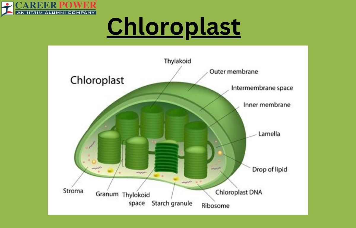
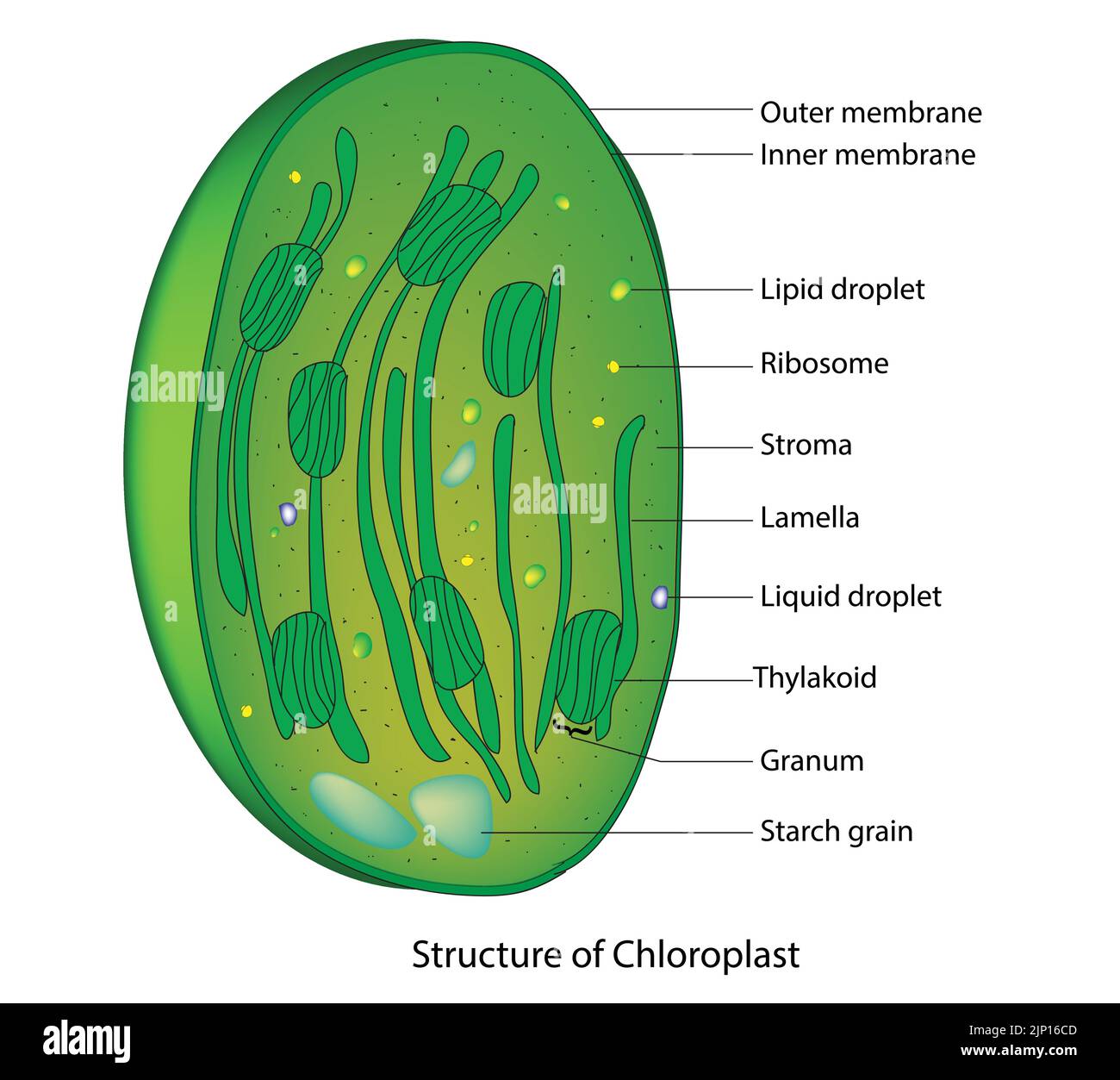
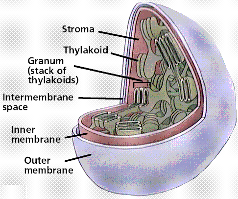
Chloroplast: Definition, Diagram, Functions and Structure

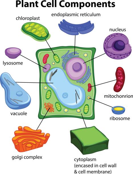
File:Simple diagram of plant cell (en).svg - Simple English Wikipedia, the free encyclopedia
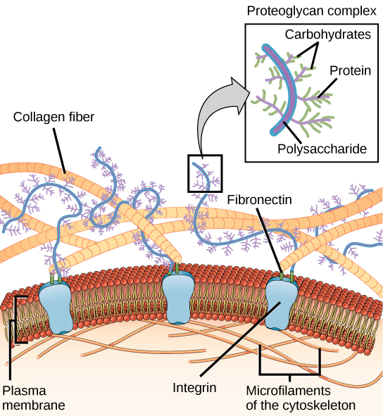
3.3 Eukaryotic Cells – Concepts of Biology – 1st Canadian Edition
Chloroplast structure hi-res stock photography and images - Alamy
media./id/840738064/vector/chart-sh

Leaf & Chloroplast Structure Botany, Plant science, Teaching biology

Animal Cells versus Plant Cells

PDF) Systematics of naviculoid diatoms (Bacillariophyta): A preliminary analysis of protoplast and frustule characters for family and order level classification
CELLS II: CELLULAR ORGANIZATION