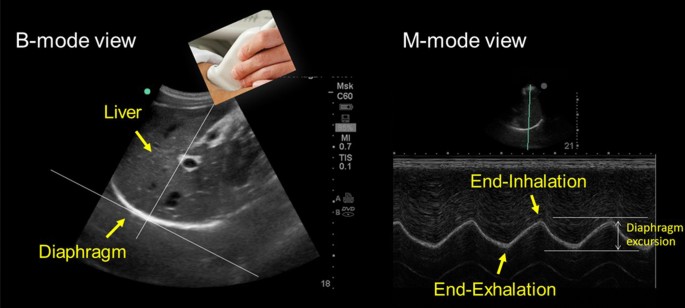
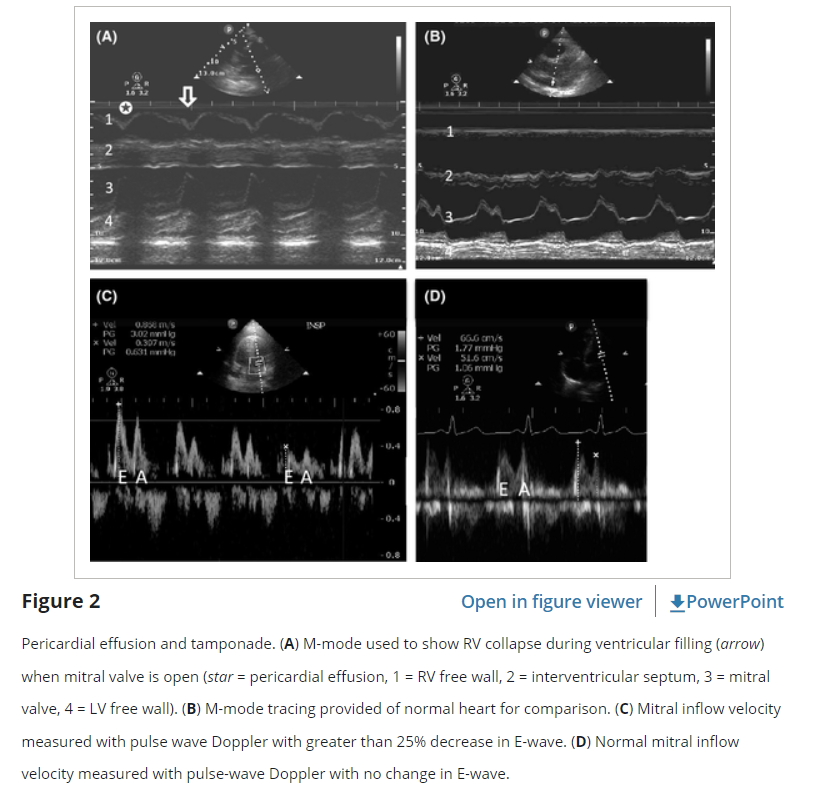
Visual estimation of IVC collapse on B-mode (grey scale image) is generally preferred to M-mode, though in theory, M-mode measurement might be able to give accurate collapsibility index. There are several reasons for this. A major limitation of IVC M-mode is that the vessel moves mediolaterally and craniocaudally during respiration, with collapse occurring off axis…

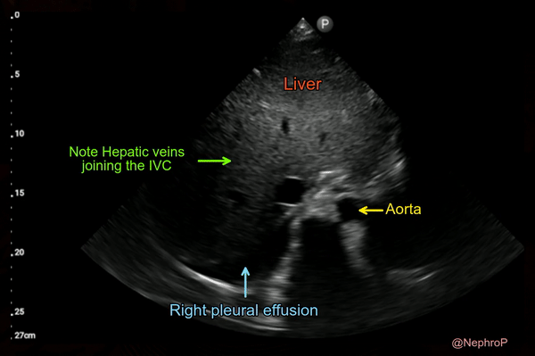
Subcostal longitudinal view of the inferior vena cava with

An intriguing tale of B-lines in a dialysis patient – NephroPOCUS

Point-of-care ultrasound in pediatric nephrology

Venous Excess Doppler Ultrasound for the Nephrologist: Pearls and Pitfalls - ScienceDirect

PDF) Venous Excess Doppler Ultrasound for the Nephrologist: Pearls
Links To And Excerpts From The First E - Effusion From The 5Es
Inferior Vena Cava POCUS: The Basics of Image Acquisition - Renal

Thread by @NephroP on Thread Reader App – Thread Reader App

Figure 13 from Congenital absence of inferior vena cava

Venous Excess Doppler Ultrasound for the Nephrologist: Pearls and

PDF) Diagnosis of Fluid Overload: From Conventional to