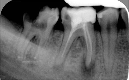
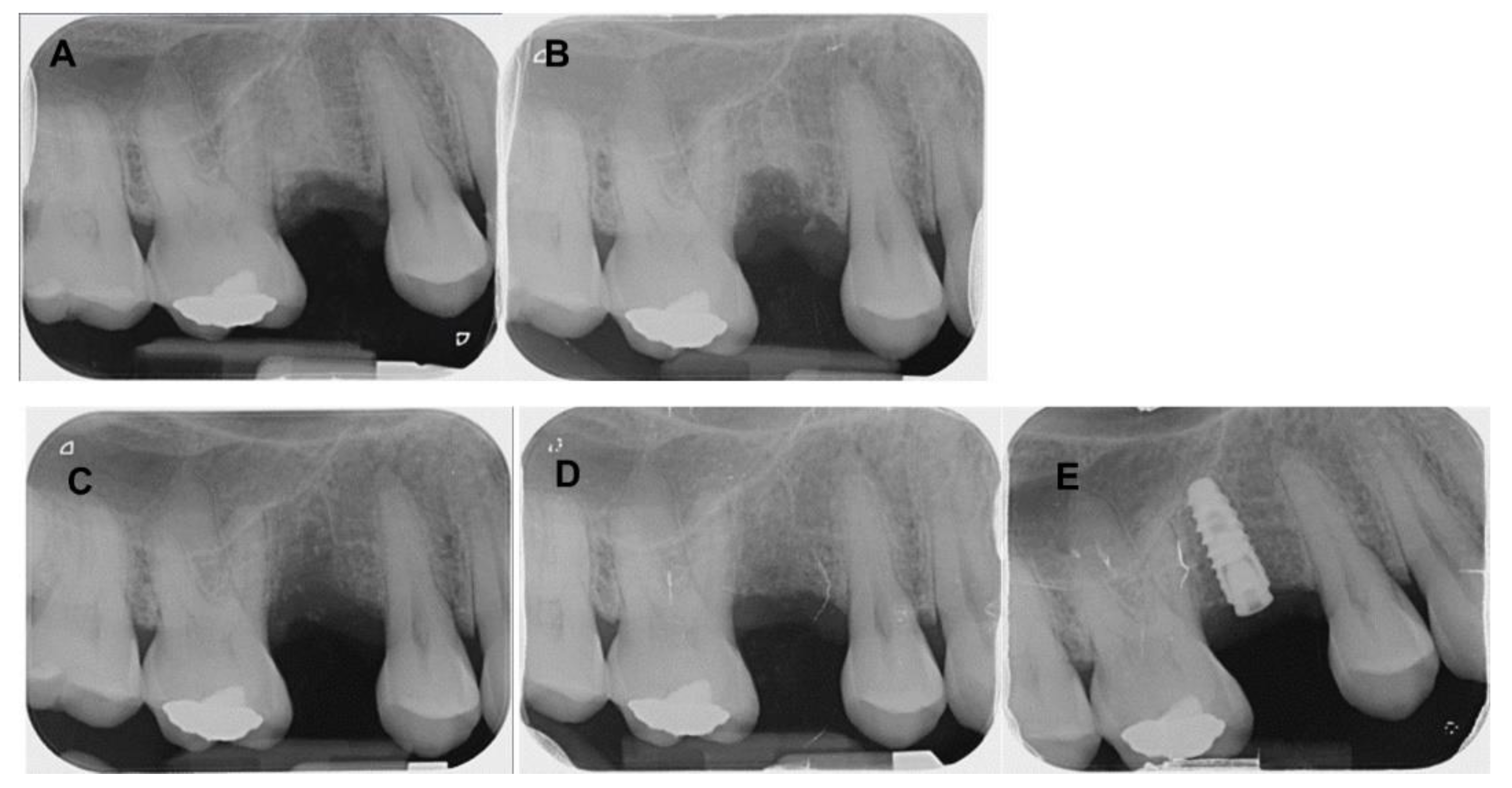
A) Preoperative intraoral periapical (IOPA) radiograph of 36. B) Post operative (IOPA) radiograph of 36. C) 1 month follow up IOPA radiograph of 36. D) 6 months follow up IOPA radiograph of 36. E) 1 year follow up IOPA radiograph of 36. - IP Indian J Conserv Endod - clinical and preclinical conservative /restorative de
6 Anatomy Pocket Dentistry

A) Preoperative intraoral periapical (IOPA) radiograph of 36. B

a) Preoperative IOPA radiograph of tooth #36. (b) Intraoral image
Cureus Graftless Immediate Dual Implant Anatomic Placement With

PDF) Direct pulp capping with bioactive materials – A case series
Coatings, Free Full-Text

Direct pulp capping with bioactive materials – A case series - IJCE

A) Preoperative intraoral periapical (IOPA) radiograph of 36. B

Preoperative IOPA radiograph showing deep caries of lower 2nd
Dentistry Journal, Free Full-Text

PDF) Direct pulp capping with bioactive materials – A case series

Paromita MAZUMDAR, Head of Faculty
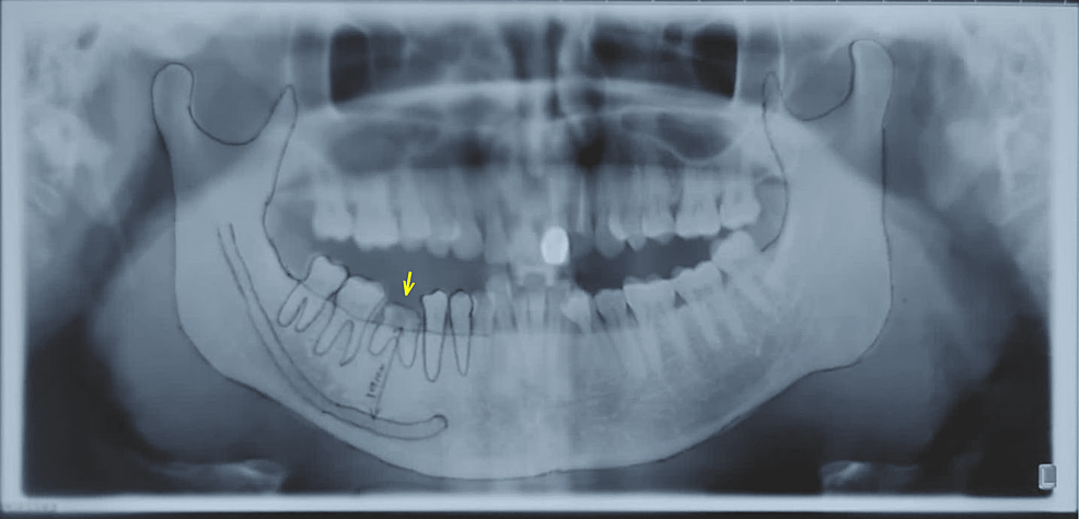
Radiograph of tooth 36: A. Ortopantomography B. Preoperative